Anencephaly – A Lethal Fetal Neurological Malformation
Jan 11 2023 | Medicover Hospitals | HyderabadThe patient of 23 years old belonging to rural Indian origin had no previous medical or surgical history, no notion of consanguinity, gravida 1, para 0, came to first trimester screening. On general examination, the patient was clinically stable, height at 157cm, weight at 60kg, no edema of the lower limbs, normal blood pressure at, 98% saturation and normal temperature.
The risk factors for this condition can be genetic, diabetics, obesity, over exposure to heat in early pregnancy and use of certain medications in pregnancy such as anti-seizure drugs.
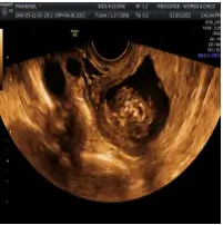
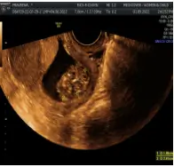
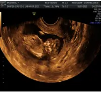
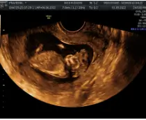
Ultrasound findings:The cranial vault, usually fully formed at 9 weeks of pregnancy, is absent. At 9–11 weeks, the cerebrovascular area can be identified, but this degenerates up to 15 weeks. Basal vessels may even be seen later. Even at the end of the first trimester, the form of the head appears unusual, and the cerebrovascular area and in particular the brain float freely, as seen on vaginal sonography. Biometric assessment at the beginning of the second trimester fails to demonstrate the bi parietal diameter, thus confirming the diagnosis. The lower part of the facial vault up to the level of the orbits is not affected, and even the brain stem remains intact. The fetal face often has a “frog-like” appearance, with prominent orbits. Myelomeningocele in the cervical or lumbosacral region may accompany this anomaly. In late pregnancy, the absence of a swallowing reflex leads to hydramnios.
