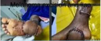
Despite its low incidence, melanoma is the most common malignant neoplasm of the foot and ankle. To achieve local control of melanoma, large surgical margins are required, thus creating important soft tissue defects. Defects located in the weight-bearing heel or in the superior mid-foot cannot be reached by conventional local flaps and may be of dimensions not suitable for local flaps. There is little evidence on the use of the medial fascio cutaneous flap based on posterior tibial perforators (Ponten flap) for this type of reconstruction.
Case Report
A 65-year-old male patient came with a history of the black-pigmented patch with central ulceration of size 4 cm in diameter involving hind foot sole skin and he was investigated by punch biopsy, which confirms malignant melanoma of foot with no other lesions in other parts of the body, without any palpable lymph nodes. He was evaluated in view of general anesthetic fitness and planned for oncoplastic surgery by a team of surgical oncologists and plastic surgeons. Intraoperatively the lesion was clearly measured and planning of wide excision with a normal margin of 2.5cm was done. The depth of excision was up to the muscle layer.
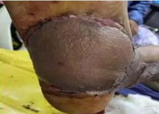
Reconstruction was planned by marking the Ponten flap cover and raised along with deep fascia from proximal to distal up to 6cm from medial malleolus, and a flap inset was given to cover the whole defect. After 3 weeks, flap division and final inset were given. After complete suture removal, the patient was advised to wear soft footwear to avoid pressure necrosis or ulceration of the flap. Even after 1 year of the follow-up period, no evidence of recurrence or pressure necrosis of the flap is observed.
Contributors
M.S., M.Ch Sr. Consultant Plastic Surgeon
Experience: 12 Years
Nellore
DR R.E.A MUTHU
M.S(General Surgery), M.Ch(Surgical Oncology) Sr. Consultant Surgical Oncologist
Experience: 5 Years
Nellore
News Letter
Medicover Hospitals Impact Newsletter August 2022